Services
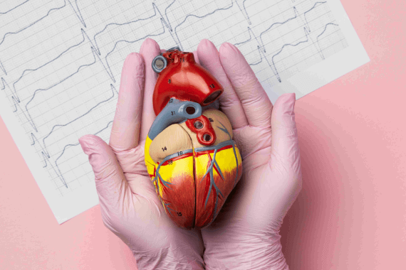
Comprehensive Cardiology Services for Your Heart's Health
Services
CARDIOVASCULAR DIAGNOSTIC SERVICES
Cardiac Catheterization (Angiography)
It allows our expert cardiologists to visualize and assess the condition of your heart's blood vessels, valves, and chambers with great precision.
Electrophysiological Studies
These studies help our expert cardiologists understand the electrical activity of your heart and identify the source of abnormal heart rhythms, also known as arrhythmias.
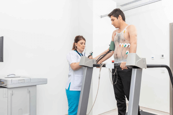
Treadmill Test
This test involves walking or running on a treadmill while your heart rate, blood pressure, and ECG (electrocardiogram) are monitored.
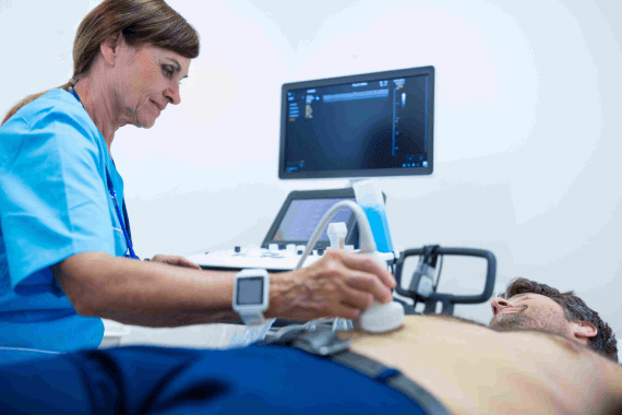
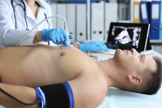
2D ECHO, Doppler, and Colour Flow Studies
These tests provide detailed and real-time images of the heart's chambers, valves, and blood vessels, aiding in the diagnosis and management of various cardiac conditions.

Stress and Dobutamine Echocardiography
These tests involve combining echocardiography with physical exercise or the administration of a medication called dobutamine to simulate the effects of exercise.
Pediatric Echocardiography
This non-invasive test uses sound waves to create detailed images of the heart, allowing our pediatric cardiologists to diagnose and monitor various heart conditions in infants, children, and adolescents.
Transesophageal Echocardiography
Unlike traditional echocardiography, TEE involves inserting a specialized probe into the esophagus to obtain closer and more precise images of the heart.
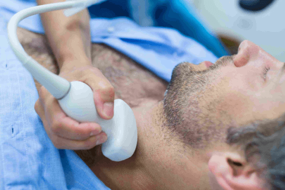
Carotid and Peripheral Doppler
This imaging test uses ultrasound technology to evaluate the health and function of these crucial arteries and vessels.
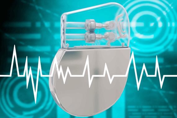
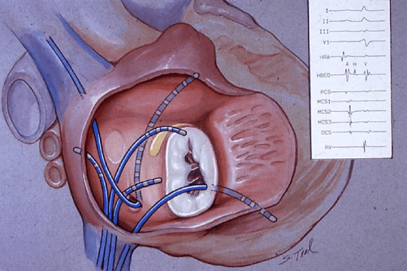
Pacemaker Analysis
Pacemakers are implantable devices used to regulate and maintain a normal heart rhythm in patients with certain cardiac conditions.
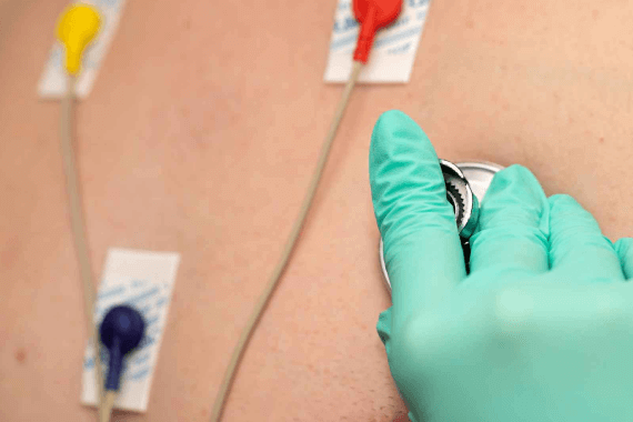
12 Leads 24 hours ambulatory Electrocardiography (Holter)
This non-invasive procedure involves wearing a portable device, called a Holter monitor, which records the electrical activity of your heart.
CARDIOVASCULAR AND INVASIVE INTERVENTIONAL SERVICES

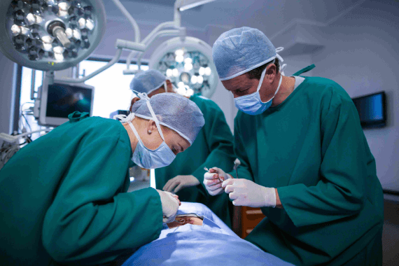
Open Heart Surgery
This procedure involves making an incision in the chest to gain direct access to the heart, allowing the surgeon to perform intricate interventions and repairs.
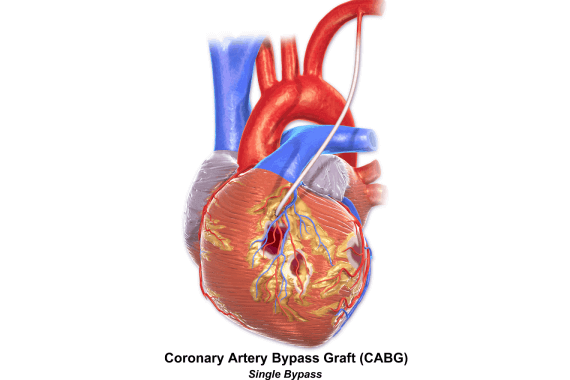
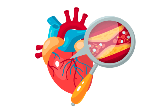
Coronary Artery Bypass Grafting (CABG)
It is commonly used to treat coronary artery disease, a condition where the coronary arteries become narrowed or blocked, reducing blood supply to the heart muscle.
Valve Replacements and Repair
The heart has four valves that regulate blood flow through the chambers, and when these valves become diseased or damaged, they may require intervention to restore proper function.

Rotational Atherectomy and Thrombosuction
These procedures target specific issues within the coronary arteries, such as plaque buildup and blood clots, to restore normal blood flow to the heart.
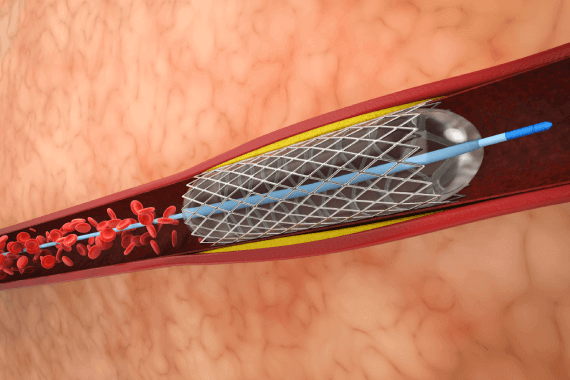
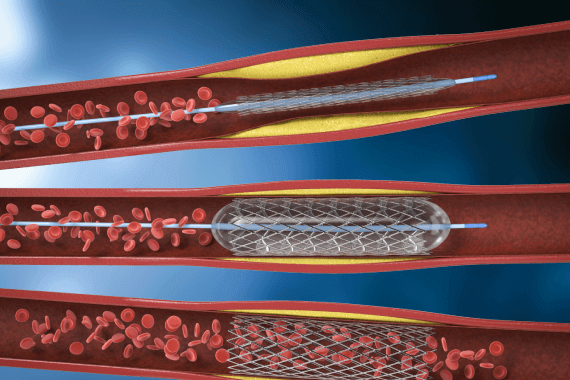
Percutaneous Transluminal Coronary Angioplasty and Stenting (PTCA)
It is commonly used to treat coronary artery disease (CAD) and relieve symptoms such as chest pain (angina) and shortness of breath.
Peripheral and Aortic Angioplasty & Stenting
These procedures aim to restore normal blood flow and alleviate symptoms caused by the reduced blood supply to the legs, arms, or other parts of the body.
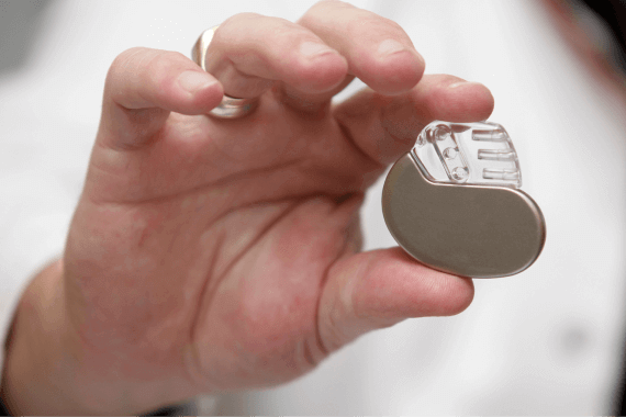
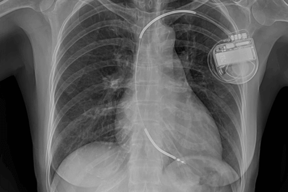
Permanent Pacemaker Implantation, Dual Chamber & BIV Pacing
These procedures involve the placement of a small electronic device, called a pacemaker, in the chest or abdomen to regulate the heart's electrical activity.
Closure of Intracardiac Defects ASD, VSD, PDA, AV Malformations
These defects involve abnormal openings or connections between the chambers or blood vessels of the heart.
Percutaneous Valvuloplasty Mitral, Pulmonary, Aortic (BMV, BPV, BAV)
Percutaneous Valvuloplasty is a minimally invasive procedure performed to treat narrowed heart valves, including the Mitral, Pulmonary, and Aortic valves.
EP STUDY AND RADIOFREQUENCY ABLATION OF ARRHYTHMIAS
These procedures are particularly effective for identifying the source of abnormal electrical signals in the heart and selectively targeting and eliminating the problematic tissue.
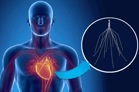
AICD and Combo Device Implantation
During the procedure, a small electronic device is implanted in the chest or abdomen. The device consists of one or more leads (thin wires) that are placed in the heart chambers or blood vessels.
IVC Filter Implantation
Pulmonary embolism occurs when blood clots from deep veins in the legs or pelvis travel to the lungs, potentially causing life-threatening complications.
Peripheral Vascular Interventions and Surgery
Peripheral Vascular Interventions and Surgery are specialized procedures offered to diagnose and treat peripheral vascular disease (PVD), a condition that affects the blood vessels outside the heart and brain.